Created by Jiří Kofránek
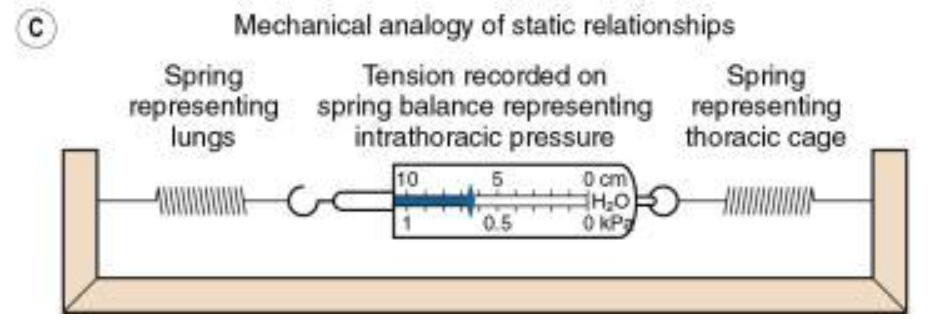
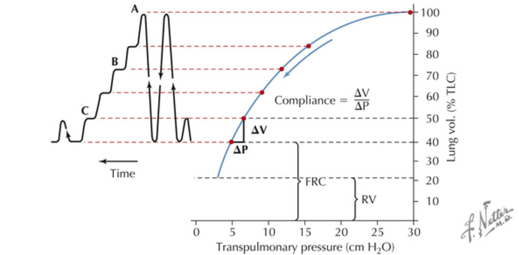
Schematic diagrams of alveoli to illustrate conditions under which static and dynamic compliance may differ. (A) Represents a theoretically ideal state in which there is a reciprocal relationship between resistance and compliance resulting in gas flow being preferentially delivered to the most compliant regions, regardless of the state of inflation. Static and dynamic compliance are equal. This situation is probably never realized even in the normal subject. (B) Illustrates a state that is typical of many patients with respiratory disease. The alveoli can conveniently be divided into fast and slow groups. The direct relationship between compliance and resistance results in inspired gas being preferentially delivered to the stiff alveoli if the rate of inflation is rapid. An end-inspiratory pause then permits redistribution from the fast alveoli to the slow alveoli.
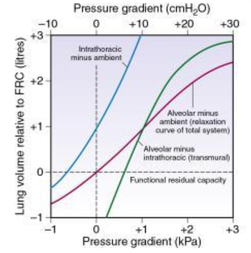
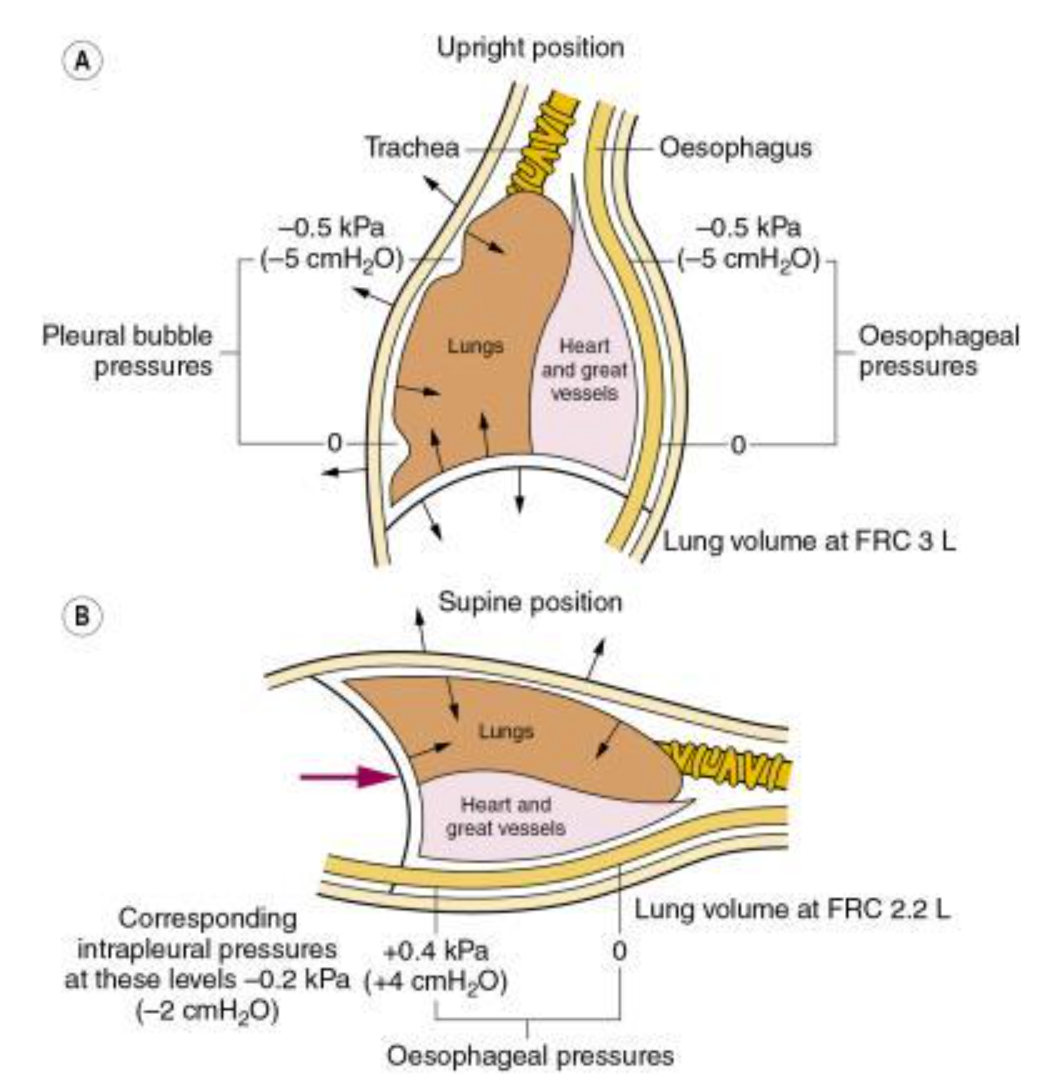
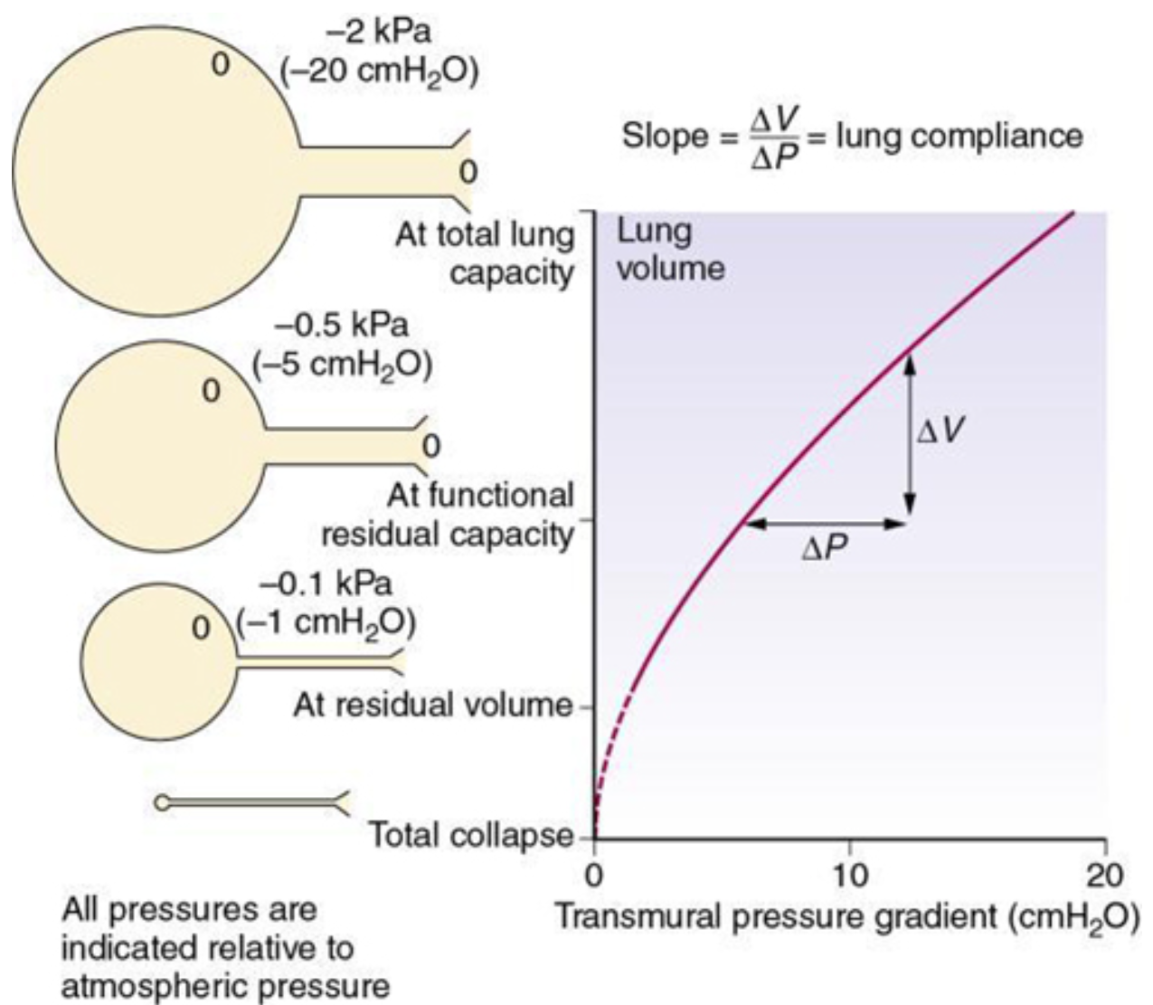
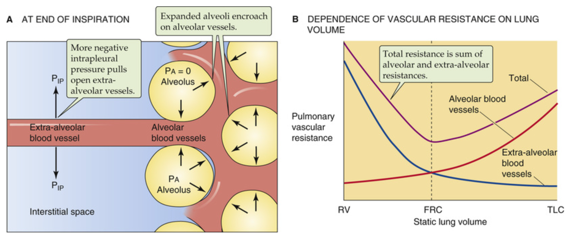
Relationship between lung volume and the difference in pressure between the alveoli and the intrathoracic space (transmural pressure gradient). The relationship is almost linear over the normal tidal volume range. The calibre of small air passages decreases in parallel with alveolar volume. Airways begin to close at the closing capacity, and there is widespread airway closure at residual volume. Values in the diagram relate to the upright position and to decreasing pressure. The opening pressure of a closed alveolus is not shown.
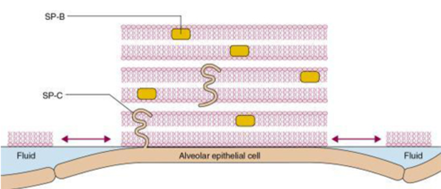
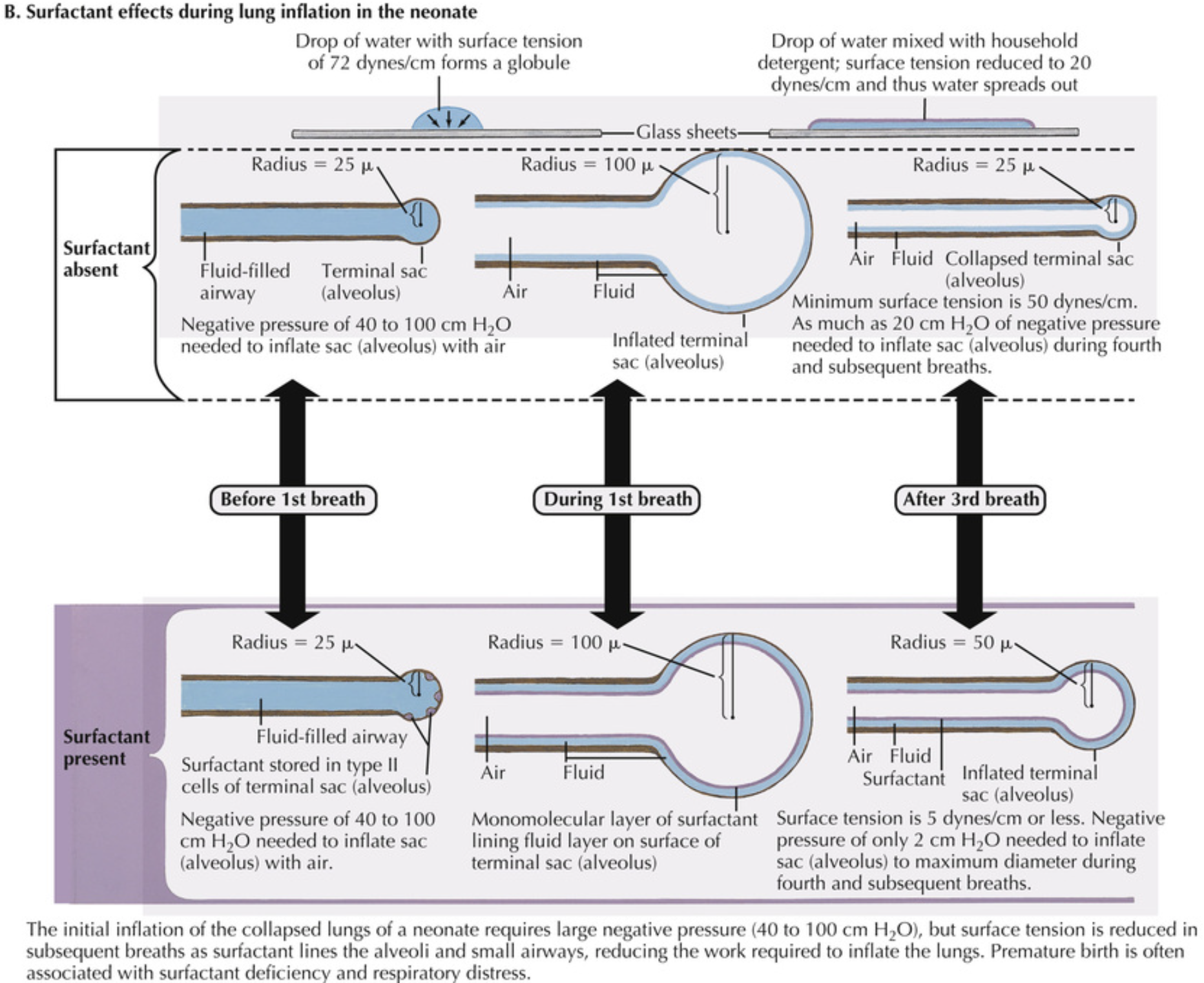
Morphological model of alveolar surfactant. Multilayered, less wettable rafts of surfactant are interspersed with fluid pools. Surfactant proteins lie within (SP-B) or across (SP-C) the lipid bilayers, facilitating the formation and dispersion of the rafts with each breath to modify the surface forces within the alveolus. Source: (From Webster NR, Galley HF. Anaesthesia Science. Oxford: Blackwell Publishing; 2006. With permission.)
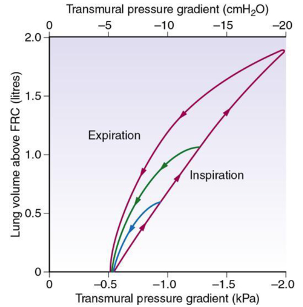
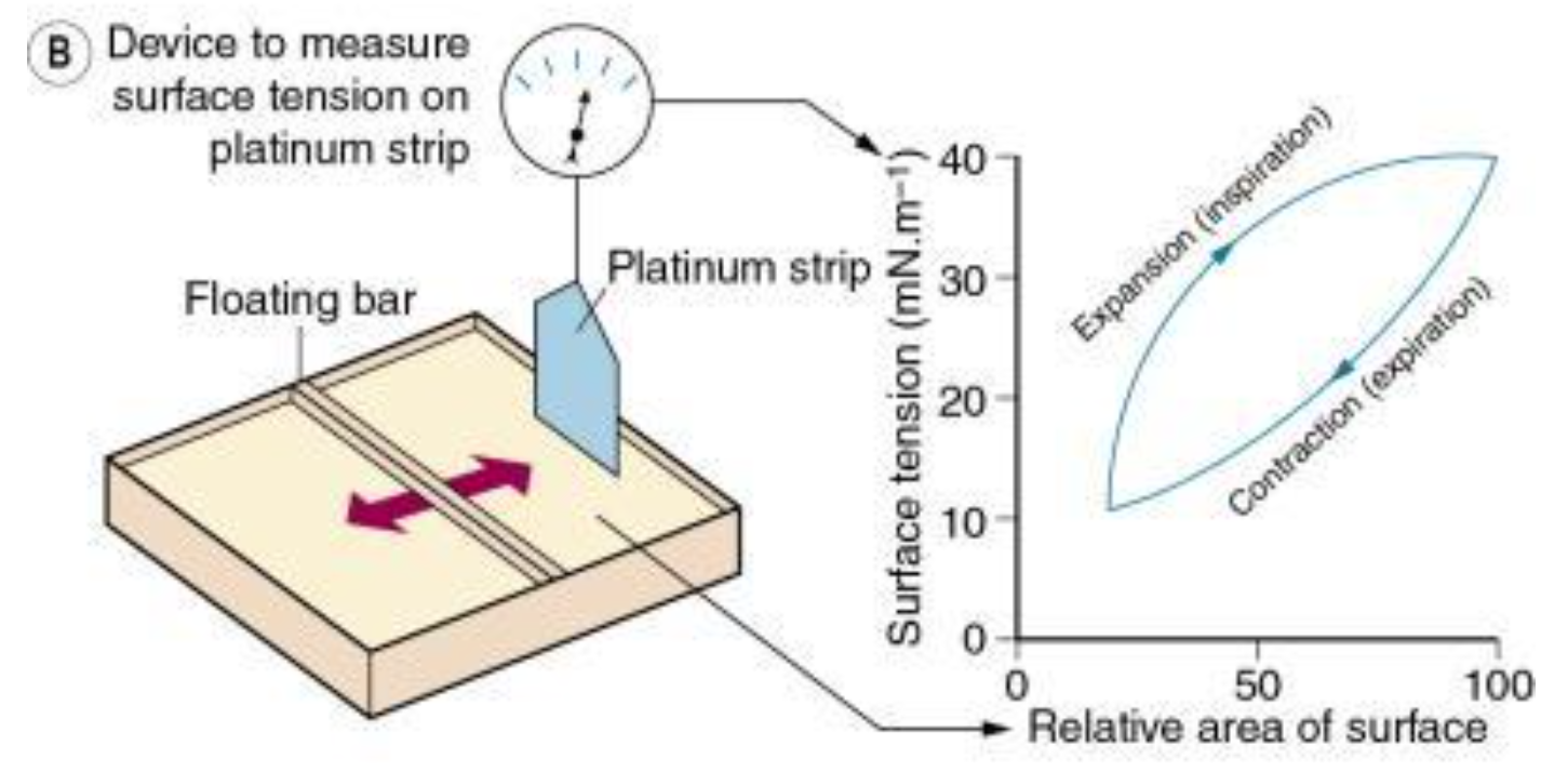
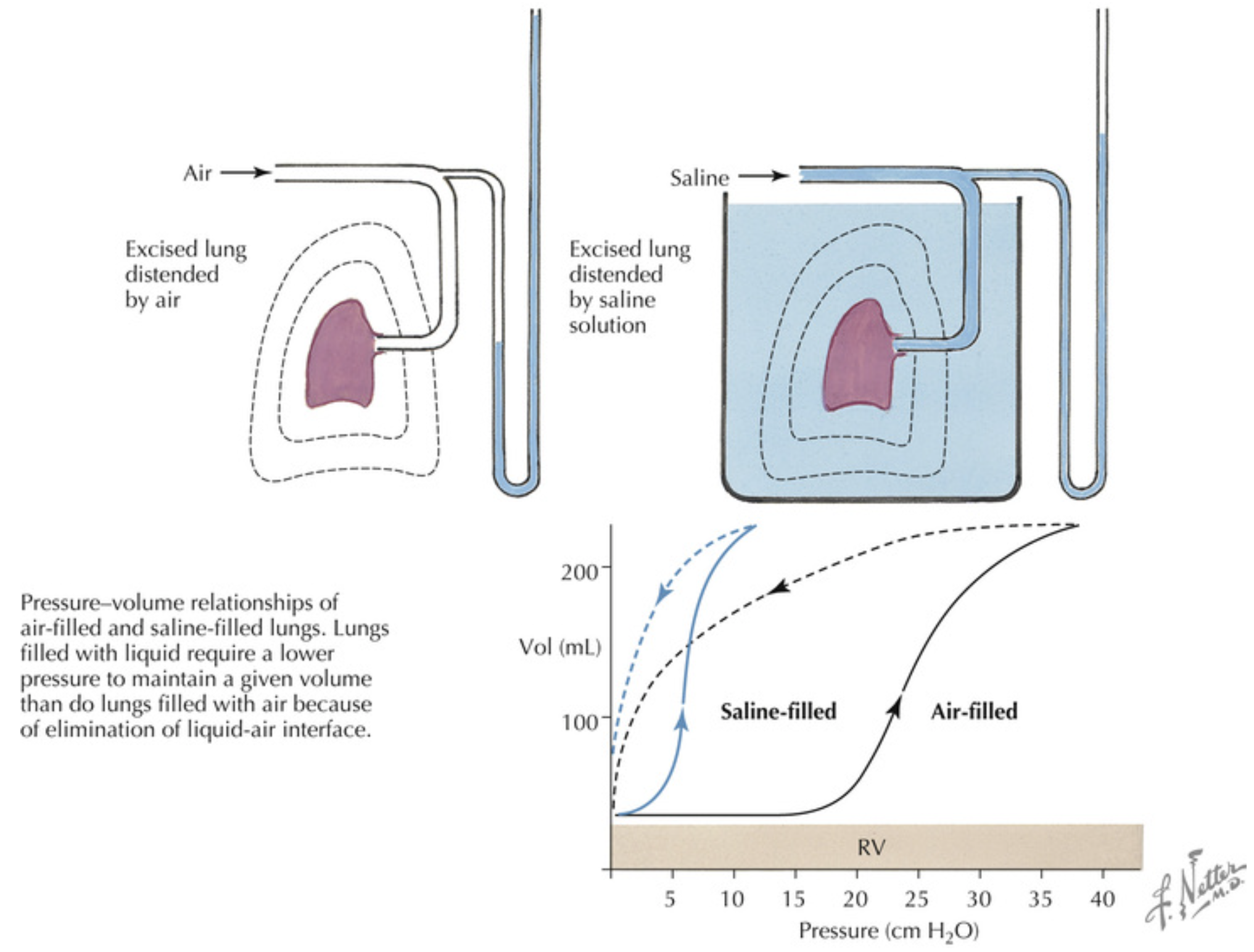
Static plot of lung volume against transmural pressure gradient (intraoesophageal pressure relative to atmospheric at zero air flow). Note that inspiratory and expiratory curves form a loop that gets wider the greater the tidal volume. These loops are typical of elastic hysteresis. For a particular lung volume, the elastic recoil of the lung during expiration is always less than the distending transmural pressure gradient required during inspiration at the same lung volume.
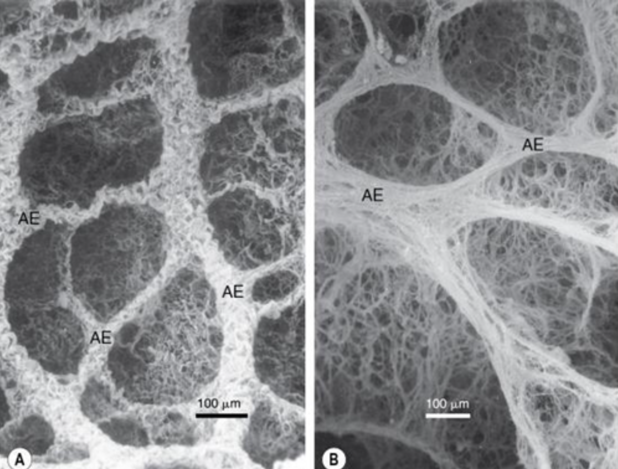
Electron micrographs of the collagen fibre network of rat lung at low lung volume (A) and when fully inflated (B).12 Note the folded, zigzag shape of the collagen at low lung volume in (A). Source: (Photograph from Professor Ohtani. Reproduced by permission of Archives of Histology and Cytology.)
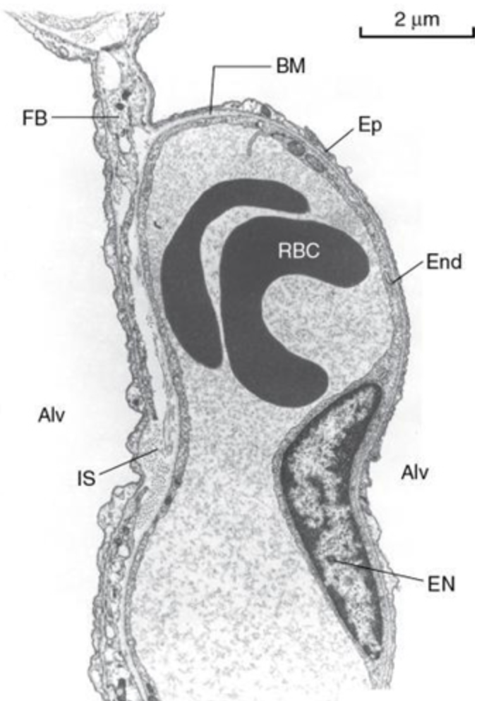
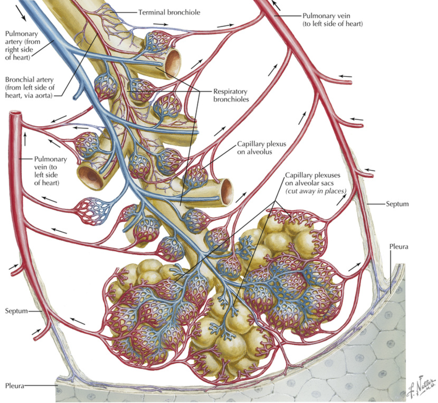
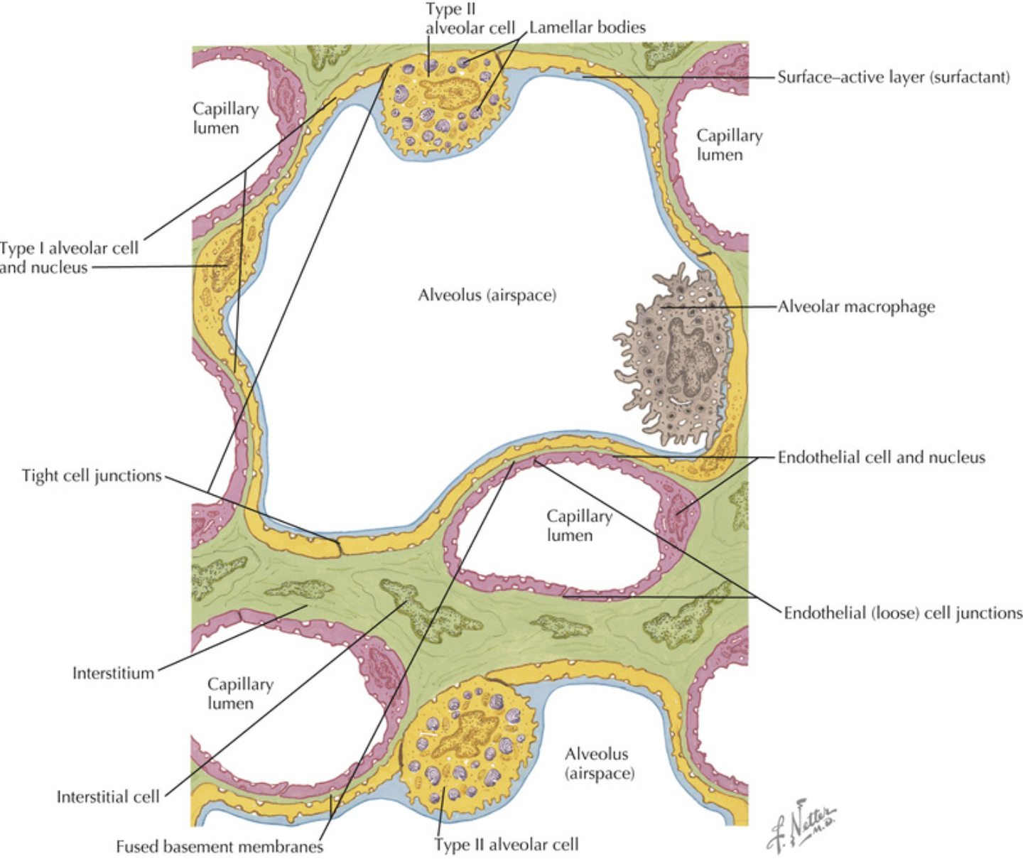
Details of the interstitial space, the capillary endothelium and the alveolar epithelium. Thickening of the interstitial space is confined to the left of the capillary (the service side), whereas the total alveolar/capillary membrane remains thin on the right (the active side), except where it is thickened by the endothelial nucleus. Alv, Alveolus; BM, basement membrane; EN, endothelial nucleus; End, endothelium; Ep, epithelium; FB, fibroblast process; IS, interstitial space; RBC, red blood cell. Source: (Electron micrograph kindly supplied by Professor E. R. Weibel.)
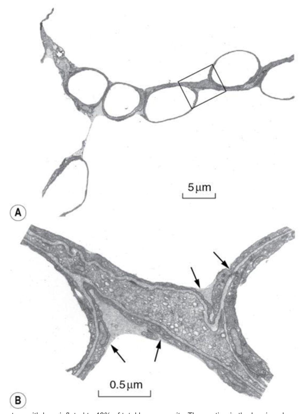
(A) Transmission electron micrograph of alveolar septum with lung inflated to 40% of total lung capacity. The section in the box is enlarged in (B) to show alveolar lining fluid, which has pooled in two concavities of the alveolar epithelium and has also spanned the pore of Kohn in (A). There is a thin film of osmiophilic material (arrows), probably surfactant, at the interface between air and the alveolar lining fluid. Source: (From reference 10 by permission of the authors and the editors of Journal of Applied Physiology.)
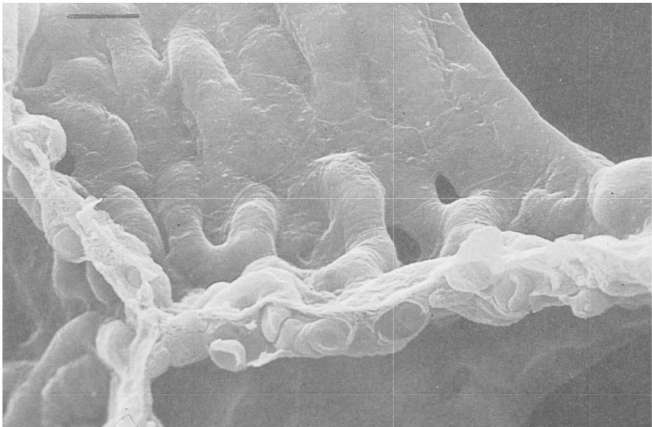
Scanning electron micrograph of an alveolar macrophage advancing to the right over epithelial type I cells. Scale bar = 3 μm. Source: (From Weibel ER. The Pathway for Oxygen. Cambridge, Mass.: Harvard University Press; 1984. With permission. © Harvard University Press.)
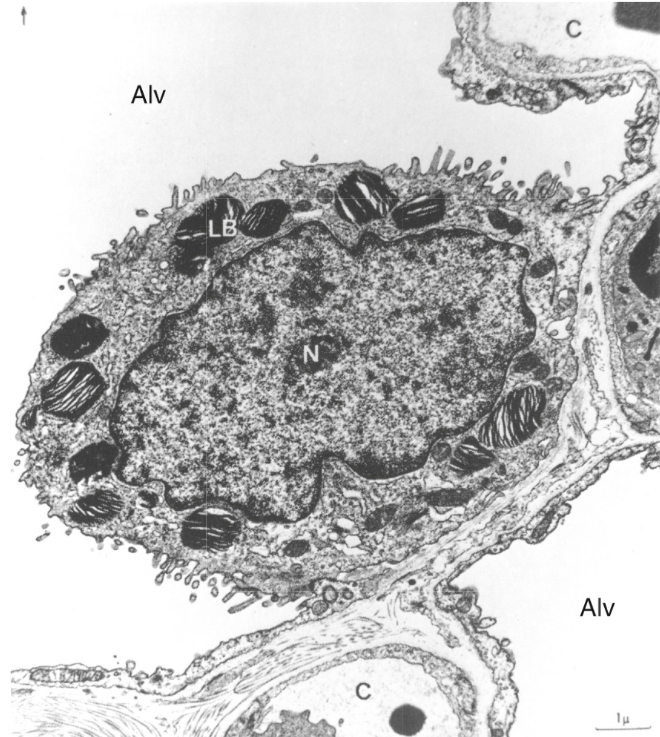
Electron micrograph of a type II alveolar epithelial cell of a dog. Note the large nucleus, the microvilli and the osmiophilic lamellar bodies thought to release surfactant. Alv, alveolus; C, capillary; LB, lamellar bodies; N, nucleus. Source: (From reference 17 by permission of Professor E. R. Weibel and the editors of Physiological Reviews.)
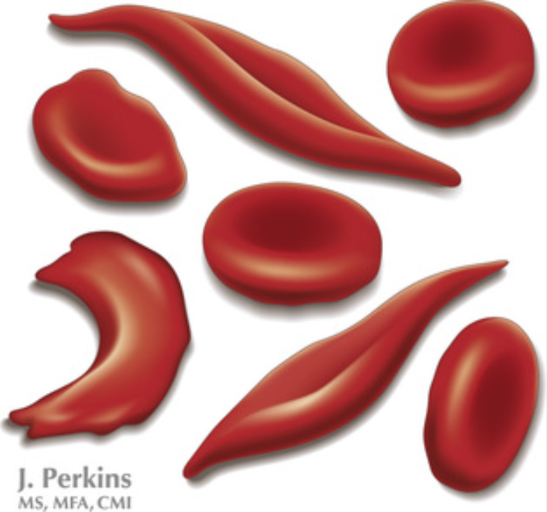
Sickled red blood cells
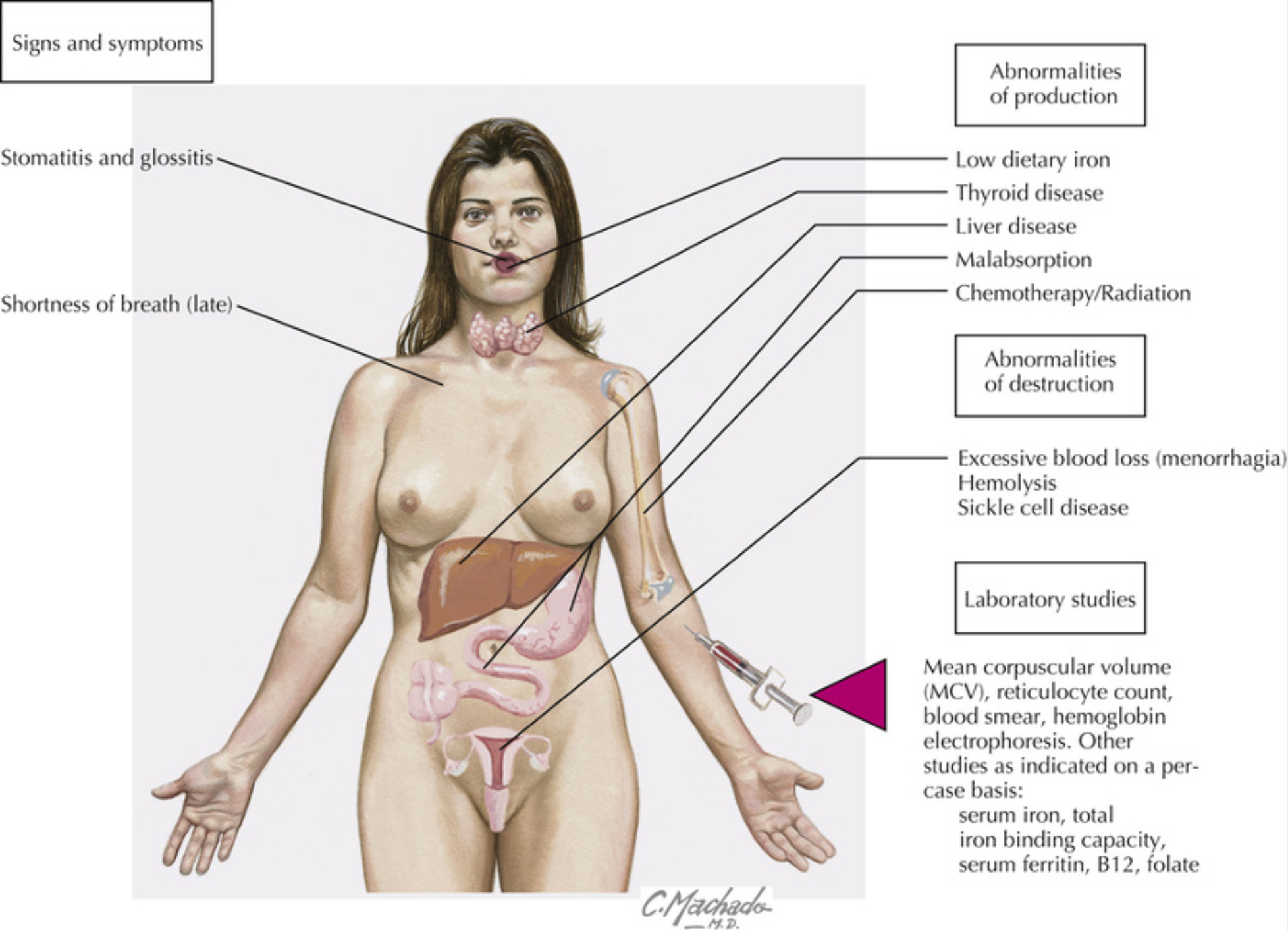
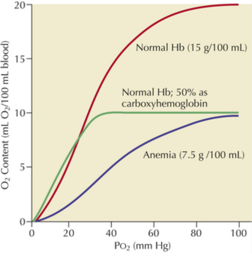
Anemia